3-D Automated Breast Ultrasound (ABUS)
It has been established that women with dense breasts are excellent candidates for supplemental screening. Automated breast ultrasound system (ABUS) is an emerging technology that has been developed to overcome the challenges of hand-held ultrasound (HHUS) and thereby to improve the availability of supplemental screening US in women with dense breasts.
ABUS is based on automated scanning of a large portion of the breast and the ability to convert 2D images to high quality multiplanar 3D reconstructed planes. This provides more reliable and reproducible imaging, with global visualization of the entirety of the breast, allowing the radiologist to interpret the data obtained by technologists. Meanwhile, virtual review of data sets enables batch reading, double reading and storage of volumes which allows to compare examinations with priors, that is expected to improve specificity in follow-up examinations and to decrease biopsy rate of benign lesions. In order to improve the utility of ABUS into screening workflow a software upgrade is needed to decrease the time necessary to acquire the acquisitions, to transmit the images and to retrieve prior ones from PACS.
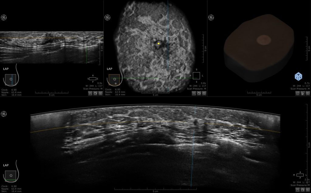
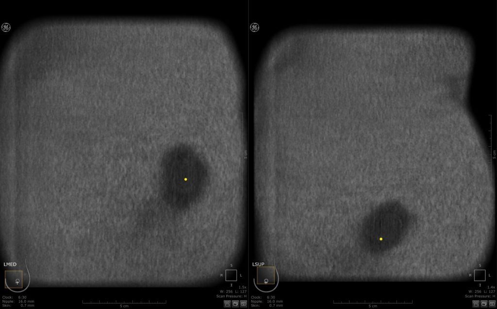
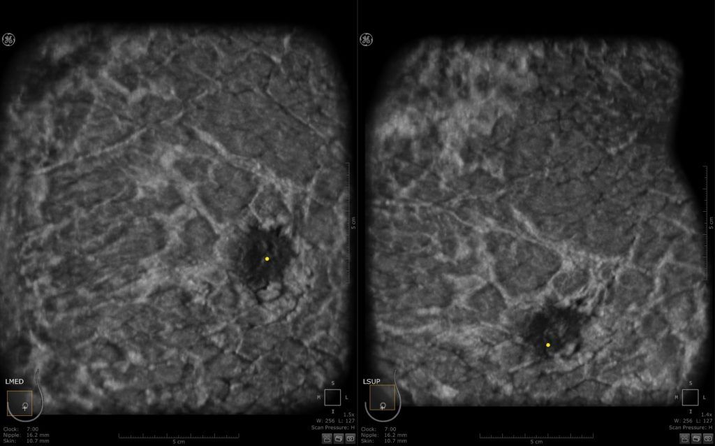
In this 43-year-old patient an irregular hypoechoic mass is seen on the left AP view in all three planes at 4:30 o’clock, 6.4 cm inferolateral to the nipple and 1 cm deep to the skin.
Normal ABUS examination in a 53-year-old-woman. Note, a large part of the breast is visualized with only one sweep.
Multiple studies that compared the performance of ABUS technology in breast cancer detection has shown similar results to handheld ultrasound (HHUS) studies and in some instances ABUS appeared to be superior to HHUS, especially in the context of architectural distortion identified in the coronal reconstruction plane.
As more data becomes available on breast density, automated breast ultrasound systems (“ABUS”) are seeing wider acceptance in breast screening. Several research articles have demonstrated it to be a significant contributor in the detection of cancers not visible on mammography or digital breast tomosynthesis (DBT). However, reviewing of ABUS cases is time-consuming. To improve reading time, a computer-aided detection (CAD) software for ABUS (“QVCAD”) was developed and has received CE mark approval. A reader study has shown this new technology has the potential to improve the reading time with no loss in diagnostic accuracy. There has been an average of 33% improved reading time among 18 radiologists in this study. These results conclude that the use of the concurrent-read QVCAD system for interpretation of screening ABUS studies of women with dense breast tissue makes interpretation significantly faster and produce non-inferior diagnostic accuracy compared to that of unaided conventional ABUS reading.
Several studies have evaluated the potential role of ABUS for breast density measurement on the basis of its three dimensional prospective, but incorporation of such techniques into clinical practice remains investigational.
Recently we explored the utility of Radiomic ABUS signature in the differentiation of benign from malignant breast lesions. This retrospective study included a total of 77 patients (all females) with biopsy proven lesions. Our results showed that the radiomic signature obtained from ABUS images is a promising tool in differentiating benign from malignant breast lesions, concluding that the automatic classification of these lesions could be successfully implemented using a radiomic signature extracted from ABUS images.
Establishment an ABUS training program for radiologists and technicians that will provide adequate training in acquiring and interpreting 3D breast volumes data obtained by ABUS will potentially improve the sensitivity, specificity and will reduce the false negative rate.
Take home points:
- ABUS is an efficient, reproducible and comprehensive technique for supplemental breast screening.
- Novel developments of ABUS will help increase the sensitivity and specificity and will lead to more accurate diagnosis of cancers.
Abbreviations:
ABUS – Three-dimensional automated breast sonography
HHUS – Hand-held ultrasound
DBT – Digital breast tomosynthesis
ABVS – Automated breast volume scanner
(UE) – Elastography US
References
- Ekpo EU, McEntee MF (2014) Measurement of breast density with digital breast tomosynthesis–a systematic review. Br J Radiol 87:20140460
- Vourtsis A, Kachulis A (2018) The performance of 3D ABUS 815 versus HHUS in the visualisation and BI-RADS characterisation 816 of breast lesions in a large cohort of 1,886 women. Eur Radiol 28: 817 592–601.
- Yulei Jiang et al. (2017) Interpretation time using a concurrent read computer-aided detection system for automated breast ultrasound in breast cancer screening of women with dense breast tissue. AJR 2018; 211:1–10.
- Vourtsis A, Berg WA. (2018) Breast density implications and supplemental screening. European Radiology. Under Press.
Papanikolaou N. et al. (2018) The performance of Radiomic ABUS signature in the differentiation of benign from malignant breast lesions. EUSOBI annual meeting 2018, Athens Greece.


