What do I need to know about breast density and supplemental screening?
Breast density has become a challenging topic with ongoing discussions as a result of the growing recognition about breast density’s masking and risk implications. The definition of breast density is the amount of fibroglandular tissue relative to fat, which comprises approximately 40% in women between 40-74 years old.
Although having dense breasts is considered a normal condition, a pool of scientific evidence has shown that women with heterogeneously dense and extremely dense breasts have a relative risk of 2-fold and 4-fold to develop invasive breast cancer compared to women with predominantly fatty breasts.
The proposed explanations for the strong positive relationship between breast density and breast cancer risk includes: a) epithelial cell content is greater in dense tissue b) the characteristics of the microenvironment is more conducive to carcinogenesis due to the production of certain growth hormones that activates cells to divide and c) dense tissue functions as a biosensor of systemic factors that increase breast cancer risk. Due to its clinical importance breast density has been recently incorporated into Tyrer-Cuzick and Breast Cancer Surveillance Consortium risk models.
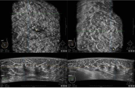
Additionally it has been widely accepted that breast density hampers the detectability of breast cancer due to its “masking effect”; therefore noncalcified cancer may be underdiagnosed because it is hidden within the breast tissue, decreasing significantly the sensitivity and specificity of mammography. This results in an increase in the interval cancer rate and a decrease in the reduction of the mortality rate.
In order to overpass the limitations of mammography an improved strategy through utilization of supplemental screening approaches has been further considered for women with dense breasts. Hand-held breast ultrasound and Automated beast ultrasound have been shown to improve the detection of invasive cancers, node negative which would be otherwise undetected in mammography due to breast density.