Breast Ultrasound and Elastography
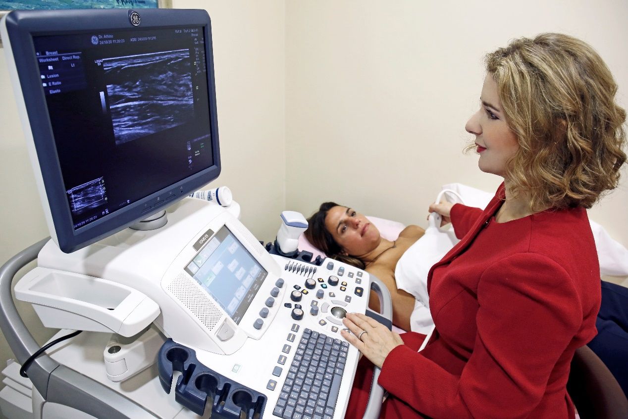
Hand-held Breast ultrasound is an imaging modality that is based on the application of sound waves and does require radiation and its related risks.
The adoption of supplemental handheld ultrasound after mammography in women with dense breasts has shown to increase breast cancer detection by 1.8–4.6 cancers per 1,000 women screened. Importantly, more than 85% of cancers found with screening ultrasound were node negative, small, invasive cancers.
During handheld breast ultrasound examination, the physician captures images from different areas of the breast, which are given to the patient along with the report. The diagnostic performance of handheld ultrasound is significantly influenced by the expertise and knowledge of the operator. In “Diagnostic Mammography Center” the examination is carried out by well experienced and trained physicians with more than 25 years expertise in this field.
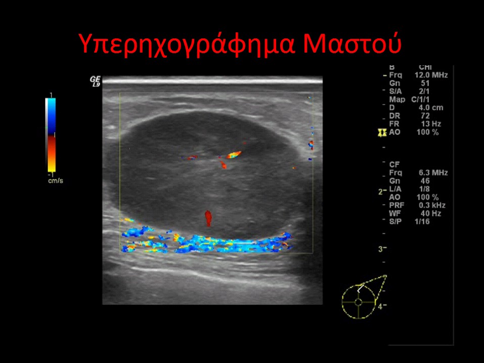
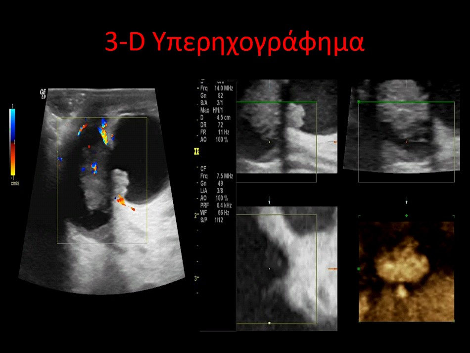
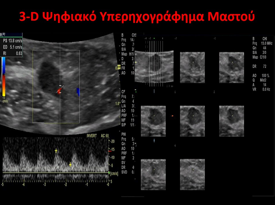
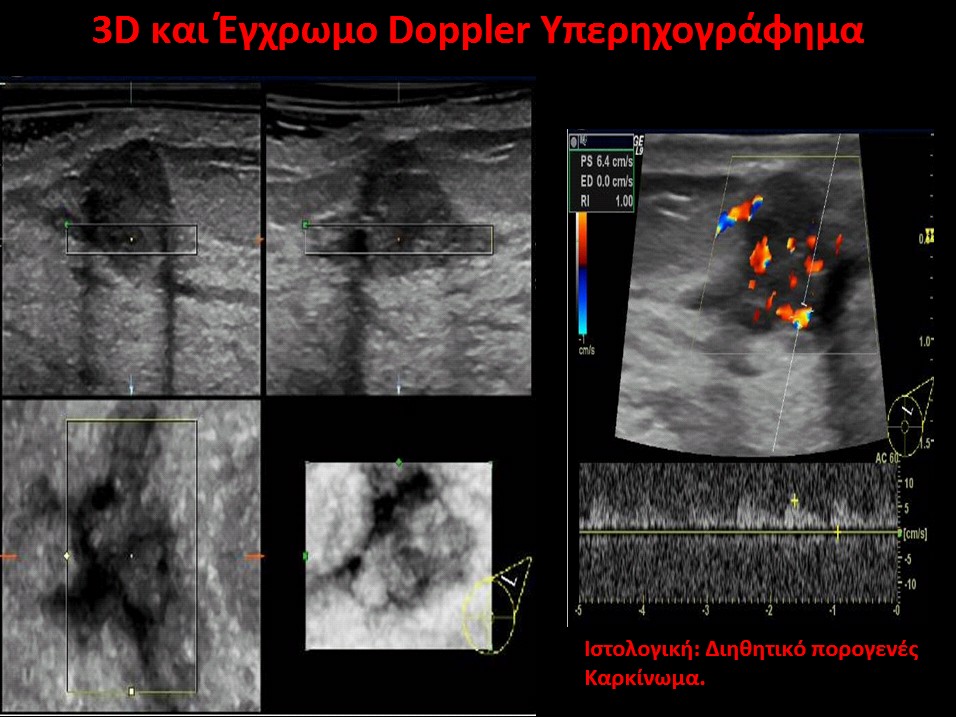
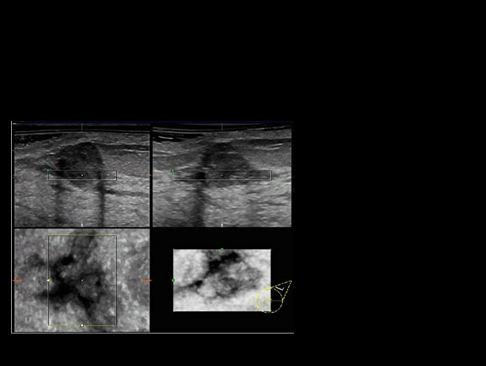
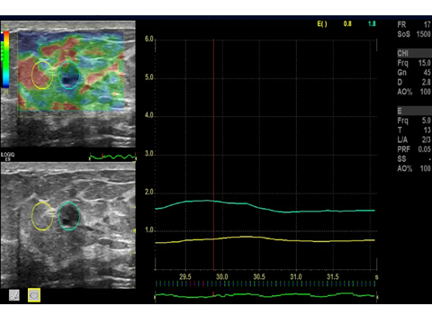
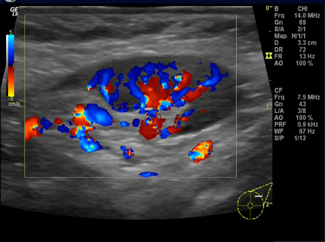
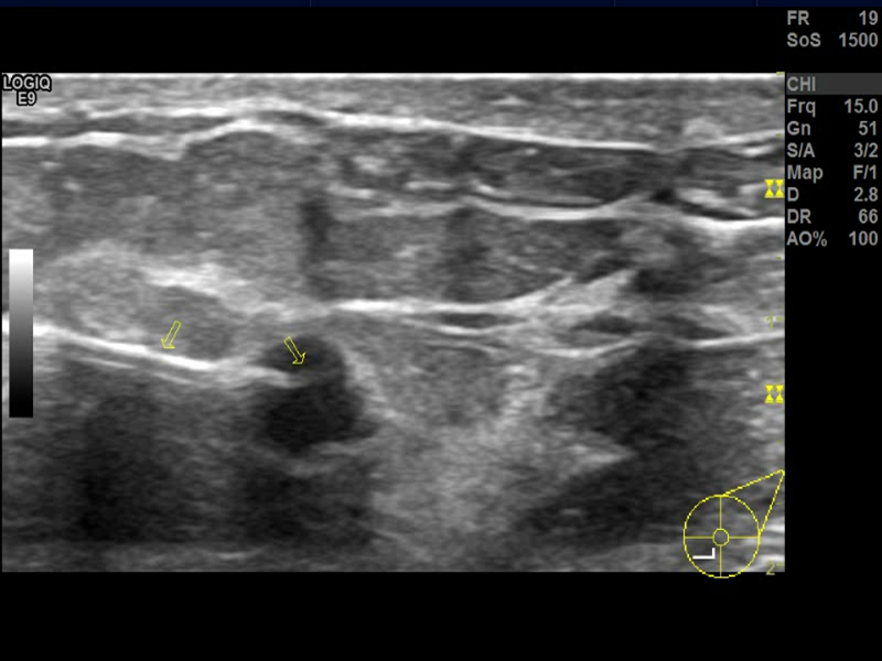
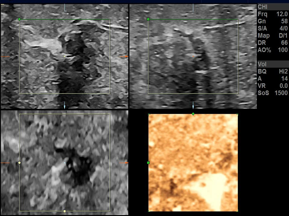
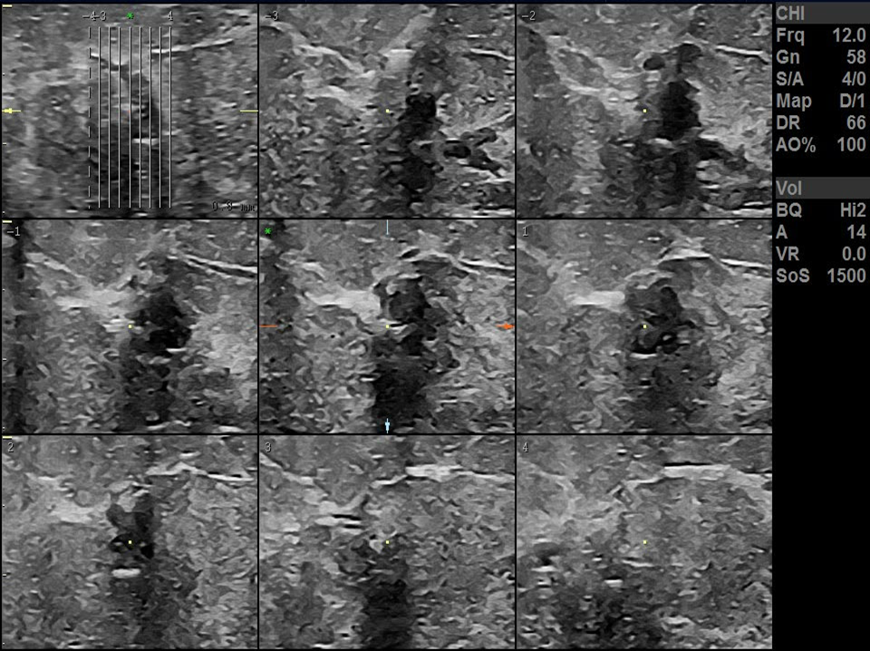
Colour-Doppler and elastography are also available on our ultrasound equipment and are used in special diagnostic cases. Color-doppler allows the identification of vascularity, which is an additional criterion used in distinguishing malignant from benign lesions. Elastography measures the tissue stiffness using colour-coded maps. This technique is based on the fact that malignant lesions are stiffer compared to benign lesions and it has shown to increase the specificity in BI-RADS characterization of breast lesions.
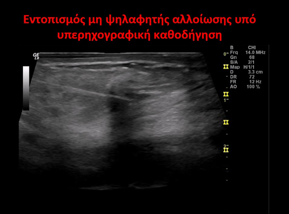
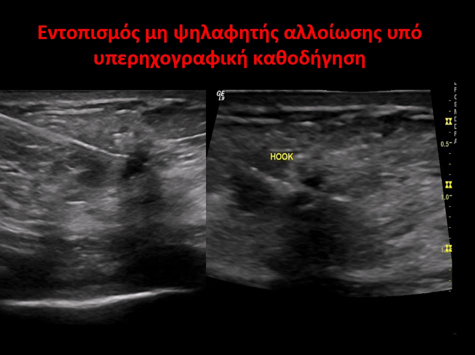
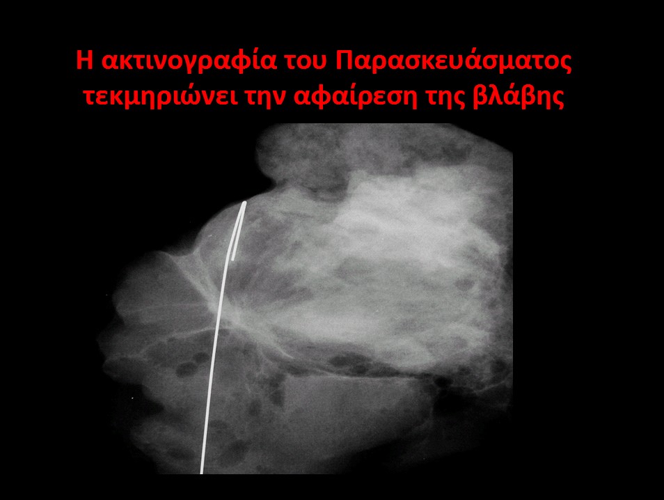
When an unexplained lesion is detected on a preventive mammogram or ultrasound, then the radiologist must proceed with the detection procedure. The “Diagnostic Mammography” center, for more than a decade, has been using the detection method. During this procedure, with the administration of a local anesthetic and the contribution of the mammogram or with the guidance of the ultrasound, the radiologist proceeds to the exact placement of the wire guide in the center of the lesion to remove it. The removed preparation is subjected to an x-ray or ultrasound to confirm that the lesion has been completely removed. According to the European Union guidelines, the radiologist is responsible for the detection process.