Artificial inteligence
The term artificial intelligence refers to the field of computer science that comprises the design of computer algorithms that mimic cognitive functions of human behavior such as learning, understanding meanings, problem solving and drawing conclusions.
The structure of artificial intelligence includes machine learning, deep learning algorithms, and deep neural networks that act similarly to the cells of the human cerebral cortex. The main feature of neural networks is the gradual improvement of algorithms through the training of the learning model, while recent technological advances have made them have a central role in our daily lives.
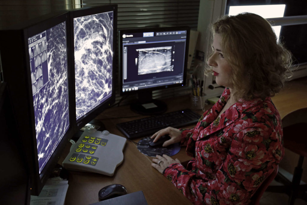
In recent years, artificial intelligence is increasingly utilized in medical imaging, predominately in mammography, tomosynthesis and ultrasound, aiding to increase diagnostic accuracy in the detection and characterization of benign from malignant lesions.
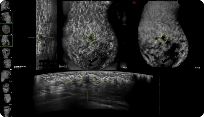
This innovative tool uses proprietary algorithms that are based on a combination of machine vision and deep learning neural network technologies that have the capability to analyze the 3D volumetric raw ABUS images and to highlight potentially significant findings. Studies have shown that the addition of CAD to interpretation of ABUS empowers Radiologist and it improves the detection of breast cancer.
In our center we have installed a leading platform of Artificial Intelligence Algorithm for Automated Breast Ultrasound (ABUS) founded by QVCAD, which was FDA approved in 2018.
Image of ABUS with the integration of CAD highlighting within the green circle a suspicious lesion.
In addition, we have installed in our center an Artificial Intelligence platform for the detection of suspicious lesions on a scale of 1 to 100, in mammography and tomosynthesis that has been developed by Transpara, Screen Point Medical. The integration of this platform in thousands of mammograms and tomosynthesis have shown an improvement in the detection of breast cancer; providing a significant assistance to the Radiologist in the fight against the disease. References:
- Priscilla J. Slanetz. Does Computer-aided Detection Help in Interpretation of Automated Breast US? Radiology 2019; 00:1–2.
- Yang S, Gao X, Liu L, et al. Performance and reading time of automated breast US with or without computer-aided detection. Radiology 2019. https://doi. org/10.1148/radiol. 2019181816.
- XuX, BaoL, TanY, ZhuL, Kong F, Wang W.1000-Case Reader Study of Radiologists’ Performance in Interpretation of Automated Breast Volume Scanner Images with a Computer-Aided Detection System. Ultrasound Med Biol 2018;44(8):1694–1702.
- Van Zelst JCM, Tan T, Clauser P, et al. Dedicated computer-aided detection software for automated 3D breast ultrasound; an efficient tool for the radiologist in supple- mental screening of women with dense breasts. Eur Radiol 2018;28(7):2996–3006.
- Kerlikowske K, Zhu W, Tosteson AN, et al. Identifying women with dense breasts at high risk for interval cancer: a cohort study. Ann Intern Med 2015;162(10):673–681.
- Alejandro Rodríguez-Ruiz, Elizabeth Krupinski, Jan-Jurre Mordang et al. Detection of Breast Cancer with Mammography: Effect of an Artificial Intelligence Support System. Radiology 2019; 00:1–10.
- Gromet M. Comparison of computer-aided detection to double reading of screening mammograms: review of 231,221 mammograms. AJR Am J Roentgenol 2008;190(4):854–859.
- Fenton JJ, Taplin SH, Carney PA, et al. Influence of computer-aided detection on performance of screening mammography. N Engl J Med 2007;356(14):1399–1409.
Lehman CD, Wellman RD, Buist DS, et al. Diagnostic accuracy of digital screening mammography with and without computer-aided detection. JAMA Intern Med 2015;175(11):1828–1837.


